Bicon Bulletin - January 2005
NEW! Bicon Instructional DVD
The new Bicon Instructional DVD contains the new Surgical Video, the revised Instrumentation Video and a comprehensive case study video. The Bicon Surgical Video includes our proven and predictable techniques that have been successfully utilized over the years by many Bicon clinicians. The Bicon Instrumentation Video is intended to provide clinicians and their staff with a detailed description and explanation for the use of various instruments of the Bicon system. The case study video presents the immediate stabilization and function surgical technique, as well as the impression, laboratory, and seating techniques for a maxillary central incisor Integrated Abutment Crown™ (IAC).
The new videos are also available online: New Surgical Video |
 |
Bicon Resorbable Collagen Membrane
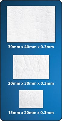 |
Guides healing of bone and surrounding tissue |
 |
 |
 |
 |
 |
 |
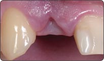
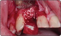
1. The appearance of a depression indicates the absence of the buccal
plate of bone.
2. Flap elevation reveals extensive loss of buccal bone and confirms the
need for bone grafting with a membrane. The height of the existing interproximal bone indicates the expected vertical level of regeneration after grafting.
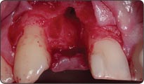
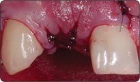
3. Bicon Resorbable Collagen Membrane is being sized and shaped to
cover the grafted site with approximately a 3.0mm margin of bone.
4. After placing the grafting material in the area of the defect, the membrane is initially tucked under the margin of the palatal flap, and then folded over the buccal aspect of the site or vice versa. Be sure that the membrane is stable and securely placed under the palatal tissue.
5. Primary closure is desirable to provide full coverage of the membrane. Dissecting and suturing the buccal aspect of the flap will allow for the coronal movement of the flap facilitating its primary closure.
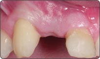
6. Six month post-operative view of the grafted site.
Bicon Shade Enhancer
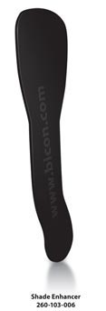 |
Facilitates efficient and accurate recording of shades
• Eliminates the frustration of taking incorrect shades. |
 |
 |
 |
 |
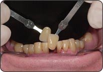
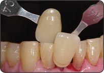
1. The wider side of the Bicon Shade Enhancer is used for recording anterior tooth shades.
2. The image is recorded with the lens perpendicular to the tooth while the shade guide is parallel to the tooth.
3. The Bicon Shade Enhancer’s thinner portion is used for the recording of shades of posterior teeth.
4. Record the image preferably at the beginning of the appointment while the tooth is still moist.
Product News
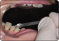
NEW! Crown Seating Tip: Designed to facilitate the tapping of an Integrated Abutment Crown™ or an extra-orally cemented crown especially in the maxillary anterior when the incisal edge of the crown is not in line with the long axis of the abutment post. The Crown Seating Tip (Part # 260-101-015) is used in conjunction with a custom thermoplastic seating jig fabricated by the technician. (See Maxillary Anterior Seating Guide below.)
|
 |
2005 Bicon Educational Opportunities & Events
As always, our calendar is replete with educational opportunities for you, your referring dentists, staff,
and laboratory technicians. Courses, lectures, events, and exhibits are frequently added, so please refer to our online calendar for
the most up-to-date information.